MRI and CT scan: What’s the Difference?
Introduction
In the realm of medical imaging, two standard diagnostic tools stand out: Magnetic Resonance Imaging (MRI) and Computed Tomography (CT) scans, which are skin tools that radiologists use. Whether you look at them from this standpoint or another, they have one thing in common – they bring us closer to understanding the human body in all of its complexity. In this one-stop shop, we’ll embark on the varied distinctions between MRI and CT scans, focusing on how they function, their inherent limitations, and their diverse applications within the medical and healthcare field.
Understanding MRI and CT scans
MRI and CT are now becoming very popular, and clinicians use technologically advanced screening methods for internal structures to diagnose a range of diseases and conditions. Regardless of similarity in an anatomical objective, these imaging modalities not only involve different technologies but also exist in different mechanisms to reach this objective.
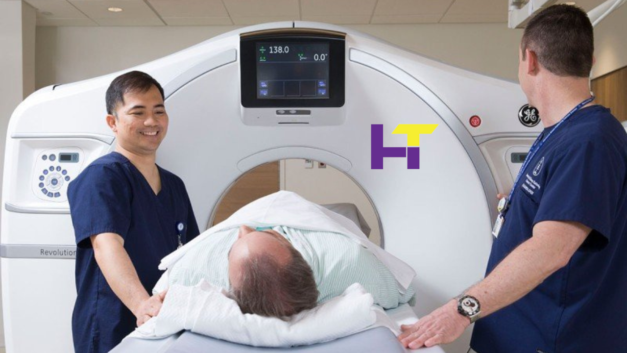
1. MRI (Magnetic Resonance Imaging):
MRI instruments are designed to produce up to a million times stronger magnetic fields than those of the natural environment. They then use radiofrequency pulses to create high-resolution images of the body’s soft tissues, which comprise muscles, organs, and nerves. CT scanners, of course, involve much higher levels of ionizing radiation than MRI, which is non-invasive while also not putting patients’ health at risk. On the contrary, MRI depends on hydrogen atoms’ behaviour inside the body, causing the magnetic fields that they are covered by.
The patient being examined during an MRI scan lies inside a large cylindrical magnet. Specific tissues are irradiated with radio waves, and special sensors pick up the signals emitted by the tissue. Such radio waves are attached to the hydrogen atoms, causing them to release signals that the receivers can pick up. Computer software will then be used to process them and create thorough cross-sectional pictures. MRI can detect and pinpoint these subtle changes due to its high contrast clarity, especially in the case of soft tissue structures like the brain, spinal cord, joints, and organs like the liver and kidney.
2. CT (Computed Tomography) Scan:
CT scans, also known as CAT scans, employ a different approach to imaging, utilizing X-rays to create detailed cross-sectional images of the body. During a CT scan, the patient lies on a motorized table that passes through a doughnut-shaped machine called a gantry. Inside the gantry, an X-ray tube rotates around the patient, emitting narrow beams of X-rays from multiple angles.
Detectors on the opposite side of the gantry capture the X-rays after they pass through the body, generating a series of two-dimensional images. A computer then processes these images to create detailed, three-dimensional representations of the internal structures. CT scans are highly effective for visualizing bones, blood vessels, and organs with high density, such as the lungs, liver, and abdomen.
Role in Pain Management
MRI and CT scans play a vital role in pain management for conditions such as sciatica, lower back pain, and shoulder pain. MRI imaging provides detailed visualization of soft tissues, aiding in the diagnosis of spinal disc herniations, nerve compressions, and rotator cuff injuries. Conversely, CT scans excel in assessing bone structures and detecting fractures, spinal abnormalities, and degenerative changes. By accurately identifying the underlying causes of pain, MRI and CT scans guide healthcare providers in formulating targeted treatment plans, including medication, physical therapy, or surgical interventions, ultimately improving patient outcomes and quality of life.
Differences Between MRI and CT Scans:
Imaging Technique:
MRI relies on the utilization of powerful magnetic fields and radiofrequency pulses to generate detailed images of soft tissues within the body. This technique offers exceptional contrast resolution and detail, making it particularly adept at visualizing organs, muscles, and nerves. In contrast, CT scans employ X-rays to produce images with superior spatial resolution, rendering them ideal for imaging dense structures like bones and blood vessels.
Radiation Exposure:
A fundamental disparity between MRI and CT scans lies in their approach to radiation exposure. MRI imaging does not involve the use of ionizing radiation, making it safe for most patients, including pregnant women and children. Conversely, CT scans expose patients to small doses of ionizing radiation, which can pose risks, particularly with repeated or unnecessary scans. Therefore, clinicians must exercise caution when determining the necessity of CT imaging and consider the potential long-term effects of radiation exposure.
Contrast Agents:
Both MRI and CT scans can utilize contrast agents to enhance image quality and highlight specific structures or abnormalities within the body. However, the type of contrast agent employed and its administration method may vary between the two modalities. In MRI, contrast agents typically consist of gadolinium-based compounds administered intravenously, which enhance the visibility of blood vessels, tumours, and areas of inflammation. In contrast, CT scans may utilize iodine-based contrast agents for intravenous administration or barium sulfate for oral ingestion, depending on the imaging objectives and patient considerations.
Image Interpretation:
Interpreting MRI and CT images necessitates distinct skill sets and expertise due to differences in image characteristics and contrast resolution. MRI images offer superb soft tissue contrast, enabling the detection of abnormalities in organs, muscles, and nerves with remarkable clarity. This makes MRI particularly advantageous for diagnosing conditions such as brain tumours, spinal cord injuries, and musculoskeletal disorders. Conversely, CT images excel in visualizing dense structures like bones, blood vessels, and areas of high density, such as tumours or haemorrhages. Consequently, CT imaging is frequently employed in trauma assessment, vascular imaging, and evaluating conditions affecting the chest and abdomen.
When to Use MRI vs CT scans:
1. MRI:
– Brain and spinal cord imaging (e.g., detecting tumours, lesions, or multiple sclerosis)
– Musculoskeletal imaging (e.g., assessing joint injuries, ligament tears, or spinal disc herniations)
– Abdominal and pelvic imaging (e.g., evaluating liver, kidney, or prostate abnormalities)
– Cardiac imaging (e.g., detecting heart abnormalities, such as myocardial infarction or congenital disabilities)
– Breast imaging (e.g., screening for breast cancer or evaluating breast implants)
2. CT scan:
– Trauma assessment (e.g., detecting fractures, internal bleeding, or organ injuries)
– Chest imaging (e.g., diagnosing lung conditions, such as pneumonia, pulmonary embolism, or lung cancer)
– Abdominal and pelvic imaging (e.g., detecting kidney stones, abdominal masses, or bowel obstructions)
– Vascular imaging (e.g., assessing blood flow, detecting aneurysms, or evaluating vascular stenosis)
– Guiding interventional procedures (e.g., biopsy guidance, drainage procedures, or tumour ablation)
Conclusion:
In summary, MRI and CT scans are invaluable tools in modern medicine, offering clinicians detailed insights into the human body’s anatomy and pathology. While both imaging modalities have their unique strengths and applications, understanding the differences between MRI and CT scans is essential for selecting the most appropriate imaging technique for each clinical scenario. By leveraging the capabilities of MRI and CT scans judiciously, healthcare professionals can make accurate diagnoses, develop effective treatment plans, and ultimately improve patient outcomes.
Read More: Caring for Your Smile: Tips and Tricks from Dentistry Experts.